

 Full blood count – how do you interpret the results and where can you have the test?
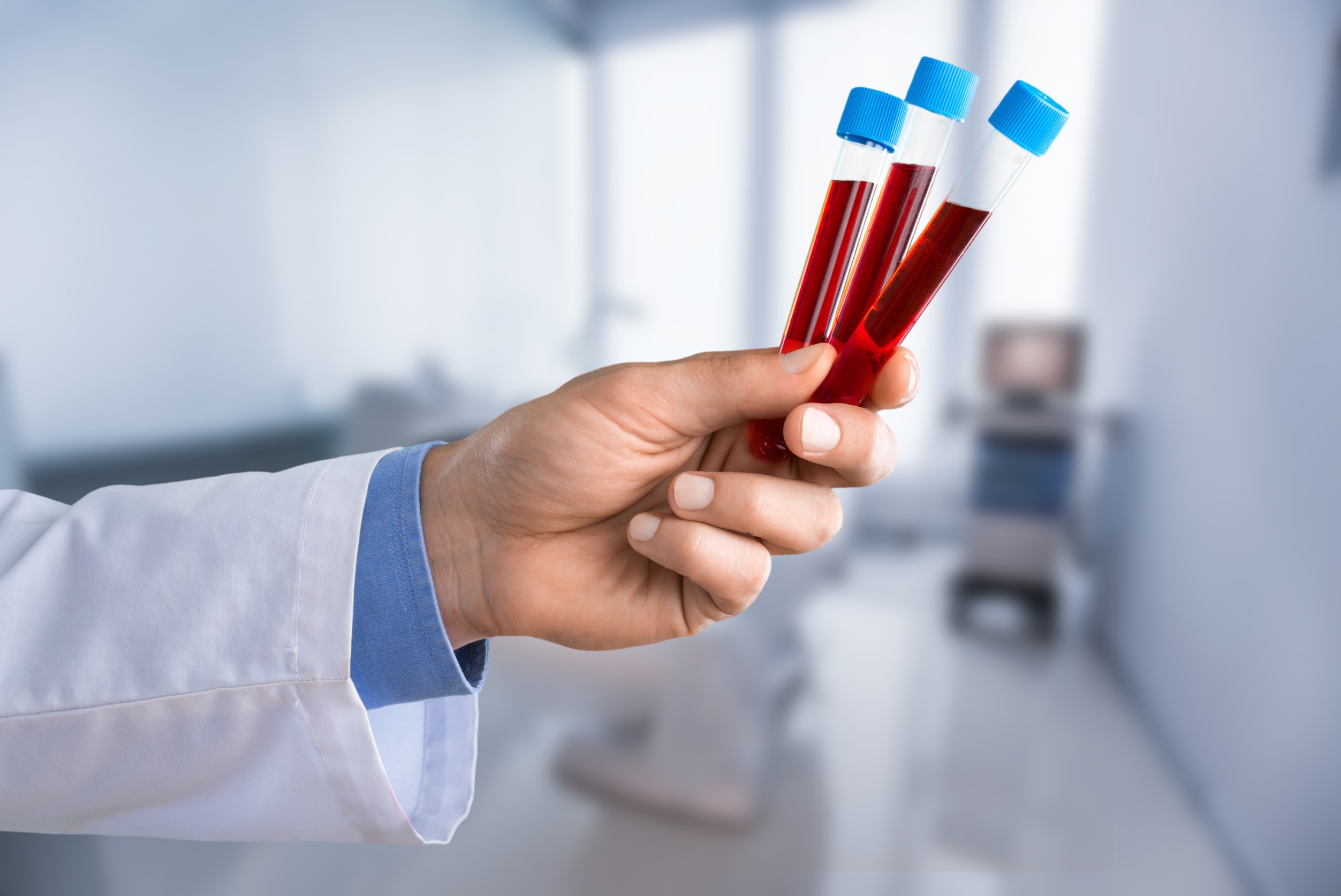
There are three types of blood cells: erythrocytes (red blood cells), white blood cells (leukocytes) and thrombocytes (platelets). Removing all the blood cells leaves the liquid part of the blood: plasma. Serum, in turn, is plasma without the components responsible for blood clotting (fibrin). Measuring the numbers of the different blood cells, as well as haematocrit and haemoglobin concentration, is known as a full blood count. A full blood count is a popular, very frequently performed test involving their quantitative and qualitative analysis.
People who feel healthy should have a blood test once a year as a preventive measure. In addition to a basic full blood count, it is advisable to have blood glucose, a lipid profile and ESR measured.
A full blood count, as well as an ESR test, is performed to check whether you have anaemia or inflammation. Remember that the earlier any abnormalities are detected, the greater the chance of a complete recovery, which is why preventive tests play such an important role. If you are experiencing pain, a full blood count and other tests help establish its cause.
You should have a full blood count on an empty stomach, because test results can change with daily fluctuations. Results may differ between the morning and the afternoon. Physical exertion or stress can also affect the result. In addition, some medicines reduce the white blood cell count. Many factors influence blood test results; you should always remember this and take it into account when analysing the tests performed. Results should always be interpreted by a doctor. The result report is issued in Polish.
PARAMETER REFERENCE RANGE WHAT A RESULT ABOVE OR BELOW THE REFERENCE RANGE MEANS
Erythrocytes (RBC – Red Blood Cell)
This test tells you the number of erythrocytes in 1 µl of blood. Erythrocytes are disc-shaped, biconcave blood cells that play a very important role: transporting life-sustaining oxygen from the lungs to all the tissues in your body, and transporting carbon dioxide from the tissues to the lungs so that it can be expelled into the air. Their production, which takes place in the bone marrow, requires a range of micronutrients and macronutrients – the most important are iron, vitamin B12 and folate (folic acid). An individual erythrocyte lives for 120 days, after which it is destroyed in the reticuloendothelial system, located in the spleen and liver.
infants – 3.8 M/µl, women – 3.9–5.6 M/µl, men – 4.5–6.5 M/µl
A result above the reference range occurs very rarely, mainly in people spending time at high altitudes in the mountains.
- o The most common cause of an increased erythrocyte count is dehydration (the blood becomes more concentrated, so erythrocytes account for a greater proportion of its volume).
- o Conditions causing chronic oxygen deficiency in the body.
Erythrocytes carry oxygen in your body, so their production increases in response to oxygen deficiency. When this occurs, the kidneys start producing more erythropoietin (EPO), a hormone that stimulates the bone marrow to produce erythrocytes. Oxygen deficiency can occur in the following situations:- o spending time at high altitudes. In high mountain areas, air pressure is lower, making gas exchange in the lung alveoli more difficult.
- o smoking cigarettes. Tobacco smoke contains carbon monoxide. Because it has a much greater ability to bind to haemoglobin than oxygen does (7 times greater), inhaling tobacco smoke causes carboxyhaemoglobin, which cannot carry oxygen, to form in place of oxygenated haemoglobin.
- o obstructive sleep apnoea (OSA) – a common condition associated with oxygen deficiency. It involves numerous long pauses in breathing (lasting even several minutes) during sleep. During these pauses, the oxygen reserves in erythrocytes are depleted, resulting in oxygen deficiency.
- o congenital heart defects in which deoxygenated (venous) blood mixes with oxygenated (arterial) blood – these include atrial or ventricular septal defects, patent ductus arteriosus, and rare conditions such as tetralogy of Fallot or transposition of the great arteries.
- o lung diseases – deoxygenated venous blood flows from the right ventricle of the heart to the lungs, where gas exchange takes place: erythrocytes release carbon dioxide (CO2) and take up oxygen (O2). Once oxygenated, the blood flows to the left ventricle of the heart and then to all your organs and tissues. Lung diseases such as chronic obstructive pulmonary disease (COPD), and the less common pulmonary fibrosis or cystic fibrosis, impair gas exchange.
- o A very rare disease that causes an increase in haematocrit is polycythaemia vera, in which erythrocyte production increases uncontrollably.
- o Tumours that secrete erythropoietin (EPO) are also an extremely rare cause of an increased haematocrit.
- o The effects of glucocorticoids during steroid treatment or in Cushing’s syndrome.
A reduced erythrocyte count indicates anaemia. It may result from blood loss or a deficiency of iron, vitamin B12 or folate (folic acid). A lower erythrocyte count also occurs in pregnant women and in kidney disease.
The following types of anaemia can be distinguished:
- o nutritional deficiency anaemia – this is the most common type of anaemia. It is most often caused by a deficiency of iron, vitamin B12 or folate (folic acid), substances needed to produce erythrocytes. The most common cause of iron deficiency is chronic bleeding; other causes include malabsorption syndromes or increased requirements (e.g. during puberty or pregnancy).
- o anaemia of chronic disease – chronic disease is the second most common cause of anaemia after iron deficiency. Various infections, cancers and autoimmune diseases activate the immune system, resulting in reduced erythrocyte production.
- o post-haemorrhagic anaemia – sudden or chronic blood loss causes fluid to move from the extracellular space into the blood vessels, diluting the blood and therefore reducing the haematocrit.
- o haemolytic anaemia – this type of anaemia is caused by abnormal, premature breakdown of erythrocytes.
- o Haemolytic anaemia is divided into congenital – where erythrocytes break down because of their abnormal structure (e.g. hereditary spherocytosis, thalassaemias) – and acquired, where erythrocytes have a normal structure but a factor causes them to break down (e.g. antibodies against erythrocytes, an artificial heart valve, infections)
- o aplastic anaemia – this occurs when the bone marrow, where erythrocytes are produced, is damaged (exposure to ionising radiation, toxic chemicals or certain medicines).
- o the body is overhydrated, although this is very rare.
Haemoglobin (HGB)
Haemoglobin is a protein contained in erythrocytes whose function is to carry oxygen molecules from the lungs to peripheral tissues. It is haemoglobin that gives erythrocytes their red colour. This test tells you how many grams of haemoglobin are present in 1 litre of peripheral blood. It is the best test for diagnosing anaemia. When deciding whether to give a blood transfusion, a doctor bases the decision on the result of this test.
A reduced haemoglobin level is generally caused by anaemia and also occurs when the body is overhydrated.
An increased haemoglobin concentration indicates polycythaemia and also occurs with disturbances of fluid and electrolyte balance (dehydration).
women – 6.8–9.3 mmol/l or 11.5–15.5 g/dl,
men – 7.4–10.5 mmol/l or 13.5–17.5 g/dl
A result above the reference range indicates dehydration.
Low values are a sign of anaemia.
Haematocrit (HCT)
This provides information on the percentage of blood made up of cellular elements, specifically erythrocytes, or red blood cells.
An increased value indicates polycythaemia or dehydration. A reduced value indicates anaemia.
children up to 15 years: 35–39%,
women: 37–47%,
men: 40–51%
MCV (Mean Corpuscular Volume), referred to as macrocytosis – the average volume of a red blood cell
80–97 fl
An increased MCV does not indicate disease, but if it exceeds 110 fl, anaemia caused by vitamin B12 or folate (folic acid) deficiency may be expected.
A reduced MCV (called microcytosis) is most often the result of iron deficiency.
MCH (Mean Corpuscular Haemoglobin)
the average amount of haemoglobin in a red blood cell; it helps determine whether erythrocytes contain a normal amount, too little or too much haemoglobin
26–32 pg
A reduced MCH indicates hypochromia (most often in iron deficiency anaemia).
MCHC (Mean Corpuscular Haemoglobin Concentration)
the average concentration of haemoglobin in a red blood cell, a measure of how fully erythrocytes are filled with haemoglobin (similar to MCH)
31–36 g/dl or 20–22 mmol/l
A reduced MCHC is typical of iron deficiency anaemia and is often seen in menstruating women.
White blood cells (WBC – White Blood Cell)
These are almost colourless, less numerous than erythrocytes and capable of movement. They live from a few days (granulocytes) to as long as 20 years (memory B lymphocytes). Their role is to protect the body against pathogens such as viruses and bacteria. All white blood cells express a molecule. White blood cells are a fundamental component of the immune system.
White blood cells are divided into:
- agranulocytes – which include:
- lymphocytes
- monocytes
- granulocytes – which include:
- neutrophils
- eosinophils
- basophils
4.1–10.9 K/µl (G/l)
An increase above the reference range signals an infection, localised or generalised inflammation, or leukaemia (increased values also occur with intense physical exertion, prolonged excessive stress, and even after extended sunbathing).
A reduced white blood cell count may be caused by a deficiency of granulocytes, lymphocytes or all cell types at the same time. It may result from bone marrow damage caused by disease or be a side effect of treatment (most anticancer medicines reduce the granulocyte count).
Lymphocytes (LYM)
These cells form part of the immune system, and their role is to defend the body against disease-causing viruses, bacteria and fungi.
0.6–4.1 K/µl; 20–45%
The lymphocyte count increases in lymphomas, chronic lymphocytic leukaemia, multiple myeloma, an overactive thyroid and childhood infectious diseases. Note: a higher lymphocyte count than in adults is normal in children up to 4 years of age!
A reduced lymphocyte count in adults may be a sign of AIDS and, to a lesser extent, other viral infections; in children, it may be congenital and require treatment as soon as possible.
Monocytes (MONO)
An increased monocyte count may be caused by infectious mononucleosis, chronic bacterial infections such as tuberculosis, syphilis, brucellosis, endocarditis and typhoid fever, protozoal infections, as well as surgical trauma, connective tissue diseases, Crohn’s disease and cancers, and may be a sign of monocytic leukaemia.
A reduced monocyte count may result from an ongoing infection or the use of certain medicines (e.g. glucocorticoids), but in medical practice it is usually not clinically significant.
140–440 K/µl (G/l)
An increase above the reference range occurs in chronic infections, after physical exertion, in iron deficiency, after removal of the spleen, during pregnancy and in essential thrombocythaemia (a neoplasm with a mild, long-term course).
A reduced platelet count may be caused by impaired production in the bone marrow (due to cancer spreading to the marrow or acute leukaemias), the effects of painkillers and antibiotics, autoimmune diseases, or destruction of platelets by bacterial toxins.
You can have a full blood count in your town or city by purchasing the test in advance from BadamySie.pl

Tests are available in the following towns and cities: Aleksandrów Łódzki near Łódź laboratory, Bełchatów blood tests, Bełżyce in the Lublin region, Bogatynia, Bychawa, Bydgoszcz, Cedry Wielkie, Chełm, Dąbrówka near Poznań, Drzewica Nowe Miasto nad Pilicą, Działdowo– Przygoda Żuromin, Garwolin, Gdańsk, Gdynia, Gliwice, Głogów, Goleniów, Gorzów Wielkopolski, Gorzyce near Wodzisław Śląski, Iłża, Jarosław near Rzeszów, Jelenia Góra, Koło, Kołobrzeg, Kostrzyn, Koszalin, Kościan near Poznań, Kościerzyna, Krakow city centre and surrounding areas, Krzywiń near Poznań, Kutno near Łódź, Kwidzyn, Legionowo near Warsaw, Lesko near Sanok, Lidzbark Welski, Lublin, Łomża near Ostrołęka, Łowicz near Łódź, Łódź, Mielec, Mińsk Mazowiecki, Nowe Miasto nad Pilicą, Nowy Sącz, Oborniki Śląskie near Wrocław, Olsztyn, Opole, Ostrołęka, Otwock, Płońsk, Poddębice, Poznań, Prabuty, Pułtusk, Raciąż near Płock, Racibórz near Wodzisław Śląski, Radom, Rogowiec near Bełchatów, Rybnik – Niedobczyce near Wodzisław Śląski, Rybnik – Niewiadom near Wodzisław Śląski, Rzeszów, Sanok, Siedlce, Sierpc, Skórzewo, Sochaczew, Szczecin, Szydłowiec, Świecie, Tuchola, Warsaw, Wodzisław Śląski, Wejherowo, Wrocław, Wysokie, Mazowieckie near Ostrołęka, Ząbki, Zabrze near Wodzisław Śląski, Zambrów near Ostrołęka, Żuromin, Żychlin near Płock
It is worth considering your full blood count result alongside your symptoms: anaemia and a raised white blood cell count slow healing and may indicate infection. Which tests to have when a wound has not healed for more than 4 weeks: A wound that will not heal: causes and tests.
Where can I get blood tests without a referral?
Blood collection centres with addresses and opening hours: Warszawa · Wrocław · Łódź · Kraków · Poznań · Gdańsk · Szczecin · Bydgoszcz · all cities.
Order your tests online at BadamySie.pl and have them done at a centre of your choice.